Figure 4.
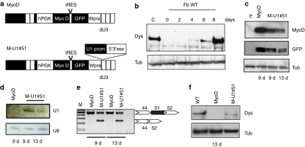
Transdifferentation of DMD Δ45–50 fibroblasts to myoblasts and dystrophin rescue upon exon 51 skipping. (a) Schematic representation of MyoD and M-U1#51 constructs. The U1 cassette expressing the 5′3′esx construct was cloned into the dU3 region. (b) Western blot analysis with anti-dystrophin (Dys) and anti-tubulin antibodies (Tub) on proteins from MyoD-infected wild-type fibroblasts (Fb WT) collected at days 0, 2, 4, 6, and 8 after induction of differentiation (C: proteins from human skeletal muscle cells). Δ45–50 fibroblasts were infected with MyoD or M-U1#51 and collected at 9 and 13 days of differentiation. Samples were analyzed by: (c) western blot, for MyoD and GFP expression, (d) northern blot, for antisense-RNA expression, (e) nested RT-PCR for exon skipping, and (f) western blot for dystrophin rescue. Western blot with anti-tubulin (Tub) antibodies was used as a loading control. cDNA, complementary DNA; DMD, Duchenne muscular dystrophy; GFP, green fluorescent protein cDNA; hPGK, human phosphoglycerate kinase promoter; IRES, internal ribosome entry site; MyoD, MyoD cDNA.

