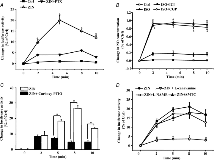
Figure 4. β2-AR regulates oxygen availability through the β2-AR–Gi–eNOS pathway.

A, C and D, cardiomyocytes expressing the MitRLuc protein were pretreated with PTX (0.5 μg ml−1, pretreated for 3 h at 37°C), carboxy-PTIO, and NOS inhibitors (pretreated for 30 min at 37°C), including l-NAME (500 nmol l−1), SMTC (100 nmol l−1) and l-canavanine (1 μmol l−1). β-AR was stimulated by different treatments, and luciferase activity was then determined as described in Methods. The data are displayed as the degree of change relative to unstimulated cells (Ctrl). B, cardiomyocytes were loaded with a DAF-2 diacetate probe, pretreated with CGP or ICI for 15 min, and then stimulated with ZIN for the indicated time. DAF-2 fluorescence was excited at 480 nm, and emitted fluorescence was measured at 540 nm. Values are expressed as the relative change compared to untreated cells (Ctrl). Single cell fluorescence was detected by laser confocal microscopy. The data represent the means of three independent experiments, and each point represents the average of ten cells.
