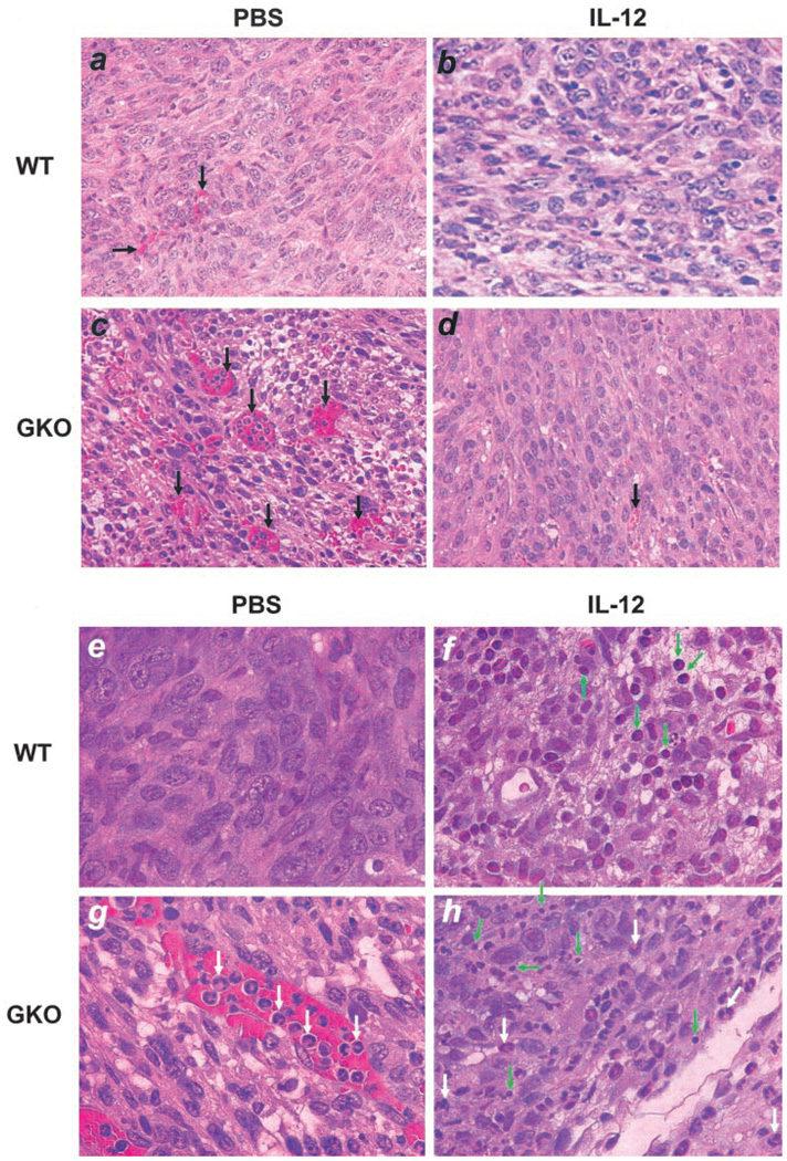
FIGURE 2.
Lymphocyte infiltration and angiogenesis in primary tumors. Paraffin-embedded tumor sections were stained with H&E and microscopically examined for blood vessels (a–d) (×200 magnification) and TILs (e–h) (×400 magnification). Shown for each group is one representative slide of three tumor samples randomly picked from the four groups. Black arrows, Blood vessels; white arrows, neutrophils; green arrows, TILs.

