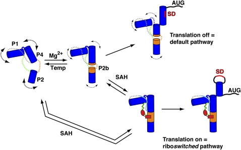
FIGURE 8.
Model of SAH riboswitch aptamer folding in Mg2+ or SAH. Blue cylinders represent the P1, P2, and P4 helices, and J4/2, J2/1, and J1/4 are shown in orange, green, and gray, respectively. In the absence of Mg2+ and SAH, the pseudoknot secondary structure is formed, the junction regions are dynamic (represented by dashed lines), and the P2b helix is not stably formed (left). In the presence of physiological magnesium concentrations (0.5–1 mM Mg2+), P2b helix formation becomes more favorable (shown as orange cylinders extending the P2 and P4 helices), but the J2/1 and J1/4 regions remain dynamic. SAH binding is supported in the presence or absence of magnesium; the structures of each are equivalent. SAH binding promotes a stable P2b helix and P2/P4 coaxial stack and the J2/1 and J1/4 regions become structured. Stabilization of the P4 helix determines the fate of the regulatory switch that either occludes or exposes the Shine-Dalgarno sequence (SD, red) for translational regulators in the absence or presence of SAH, respectively.

