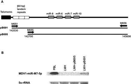
FIGURE 3.
Northern blot analysis of the role of the core promoter in mature miRNA expression. (A) Schematic diagram of the LAT-miRNA promoter and the miRNA cluster. Each black line under the representation corresponds to a DNA fragment amplified from the bacmide p-RB-1B genome and inserted into the pBS-SK vector. (B) Northern blot analysis. Each lane was loaded with 15 μg of total RNA extracted with Trizol. Peripheral blood leukocytes were recovered 21 d after infection, from highly susceptible B13 chickens infected intramuscularly with RB-1B (1000 PFU of RB-1B). RNA was extracted from infected PBL, untransfected LMH cells and LMH cells transfected with pB691 or pB695. A 32P-5′ end-labeled DNA oligonucleotide probe complementary to the miRNA was used to detect the miR-7-specific signal. Ribosomal 5s RNA (ethidium bromide staining, EB) levels were used to check for equal loading.

