
Figure 3.
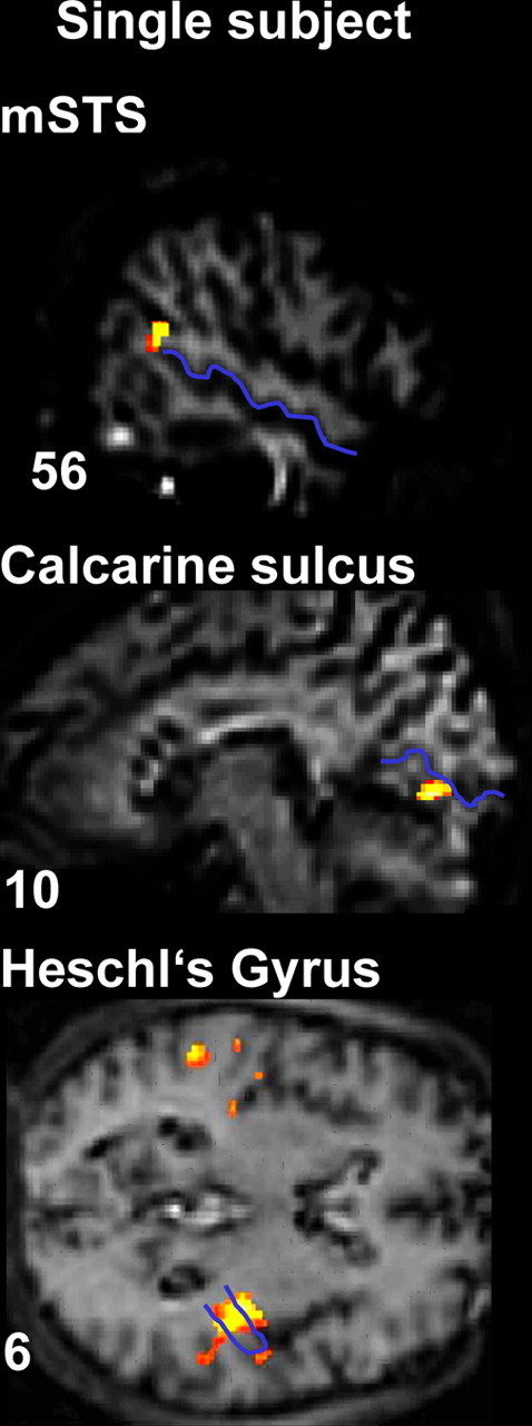
fMRI results: BOLD signal differences for corresponding minus noncorresponding audiovisual stimulation in an illustrative single subject. mSTS, visual cortex, and auditory cortex activations are shown, with the STS, the calcarine fissure, and Heschl's gyrus highlighted in blue on that individual's anatomical scan. Localization of the effects with respect to these anatomical landmarks was implemented in every individual.