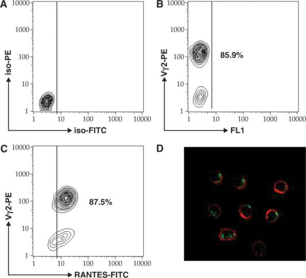
Fig. 2.
Expanded Vδ2 T cells uniformly express cytoplasmic RANTES. In vitro-expanded Vδ2 T cells were stained with PE-labeled antibodies to Vδ2 TCR chain, permeabilized, and then stained with anti-RANTES FITC-labeled antibodies. IgG isotype controls are presented a presented in panel A. More than 85% of cells were positive for Vδ2 (B) and double positive for Vδ2 and RANTES (C). A small population of RANTES single-positive cells were also detected (C). We confirmed the presence of cytoplasmic RANTES (green) in Vδ2-positive cells (red) by confocal microscopy.

