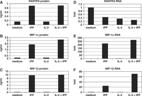
Fig. 5.
Regulation of RANTES mRNA and protein in Vγ2Vδ2 T cells. A Vγ2/Vδ2 T cell line was rested in medium supplemented with 10 U ml−1 IL-2 for 8–10 days, then washed and re-suspended in the same medium. Control cells were cultured in medium plus 10 U ml−1 IL-2. Secreted RANTES and MIP-1β levels were measured after 4 h of IPP treatment (A and B). The mRNA for RANTES was present at highest amounts prior to IPP addition and was reduced after stimulation (C). In contrast, MIP-1β mRNA was not present before IPP addition and required IPP for maximal expression (D). Cell stimulation decreased the levels of cytoplasmic RANTES and the addition of cycloheximide had little effect (E), compared with the increase in cytoplasmic MIP-1β that was sensitive to cycloheximide inhibition (F). The experiment was repeated three times using expanded Vγ2Vδ2 T cells from three different donors. The pattern of responses was identical for each Vγ2Vδ2 T cell line, but the absolute levels of chemokines varied for each donor. Accordingly, a representative experiment is shown here.

