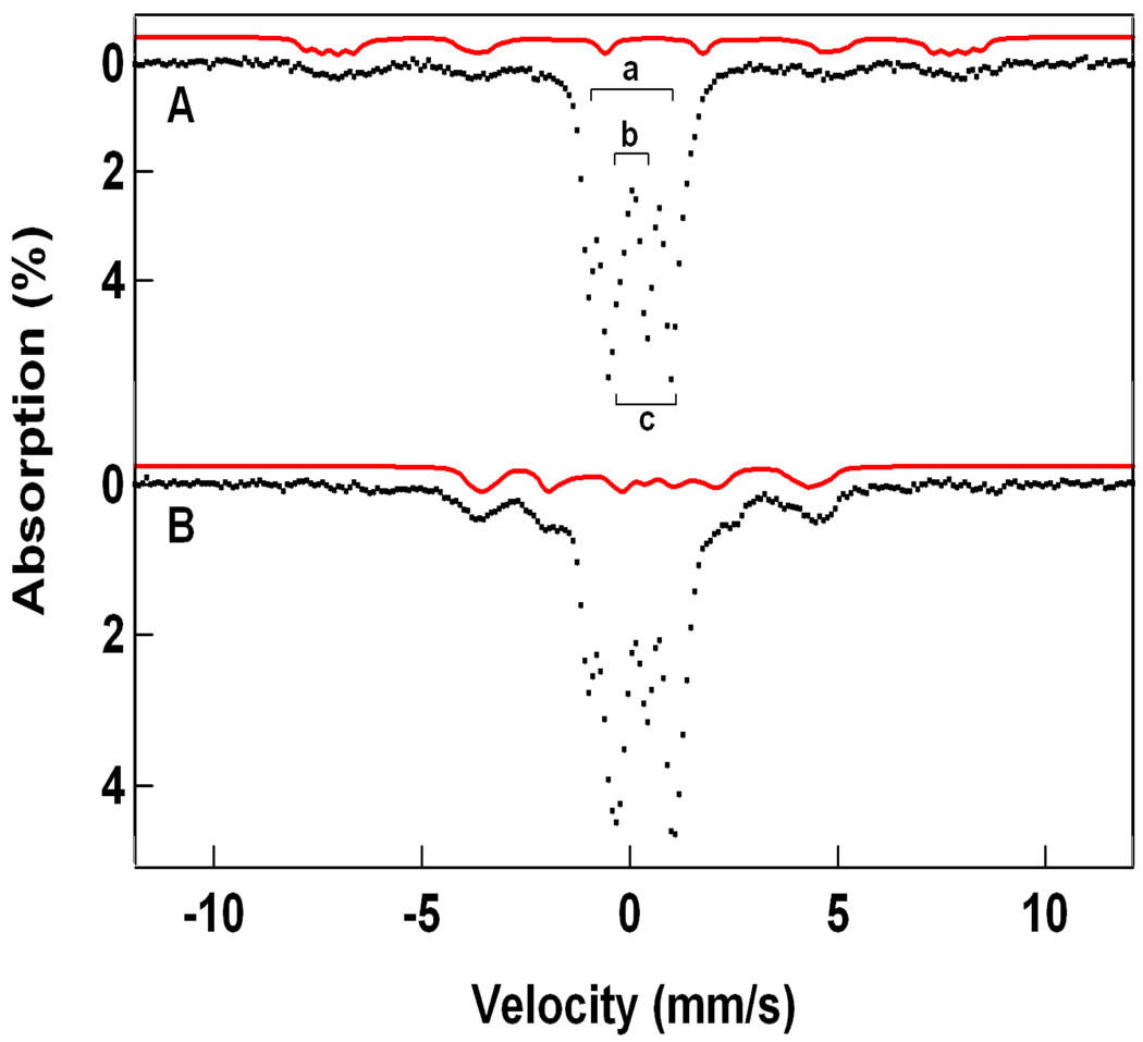
Figure 10.
Mössbauer spectra of 4 (A) in 3:1 MeOH/MeCN and 60Co irradiated 4 (B) recorded at 4.2 K in a 50 mT field applied parallel to the γ-rays. (A) The downward brackets in (A) label the FeIV sites of 4; the letter “b” designates the site with a terminal oxo group. The upward bracket marks the lines of the diferric contaminant (22% of Fe). The solid line above the spectrum is a simulated representation of a high-spin FeIII contaminant, representing ca. 10% of the Fe. The two ferric species were not affected by the irradiation with 60Co. (B) Spectrum of the sample of (A) after 60Co irradiation at 77 K. The solid red line is the simulation of 1-OH shown in Figure 7B. The high-spin FeIII contaminant has been removed using the simulation shown in (A).

