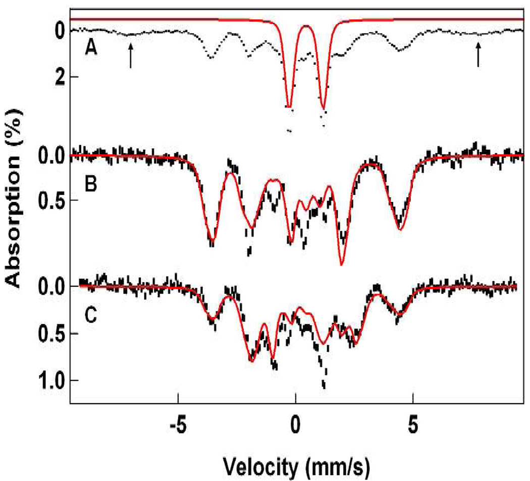
Figure 7.
Mössbauer spectra of 1-OH in 3:1 PrCN/MeCN recorded at 4.2 K in 50 mT fields applied parallel (A, B) and perpendicular (C) to the observed γ radiation. The red line in (A) outlines the spectrum of a diiron(III) contaminant; its contribution has been removed in (B) and (C). The solid lines in (B) and (C) are spectral simulations based on eq 1 using the parameters listed in Table 1. See also Figure 8. The arrows in (A) mark the outer lines of a high-spin ferric contaminant.

