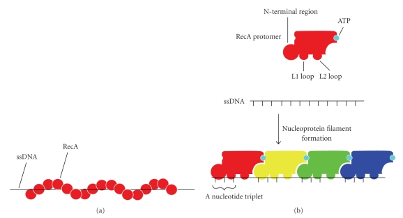
Figure 8.
A schematic illustration of RecA-ssDNA interaction in the nucleoprotein filament. (a) A schematic representation of a RecA-ssDNA nucleoprotein filament. The filament comprises a helical structure. RecA molecules are shown as red spheres and the ssDNA as a black line. (b) A schematic model of RecA-ssDNA interaction. The RecA protomer has the L1 and L2 loops and the N-terminal region to make contact with the ssDNA. The bound ssDNA comprises a nucleotide triplet with a nearly normal B-form distance between bases followed by a long internucleotide stretch before the next triplet. The ATP binds to RecA-RecA interfaces. The schematic model was prepared from the crystal structure of RecA-ssDNA complex (PDB ID: 3CMW).

