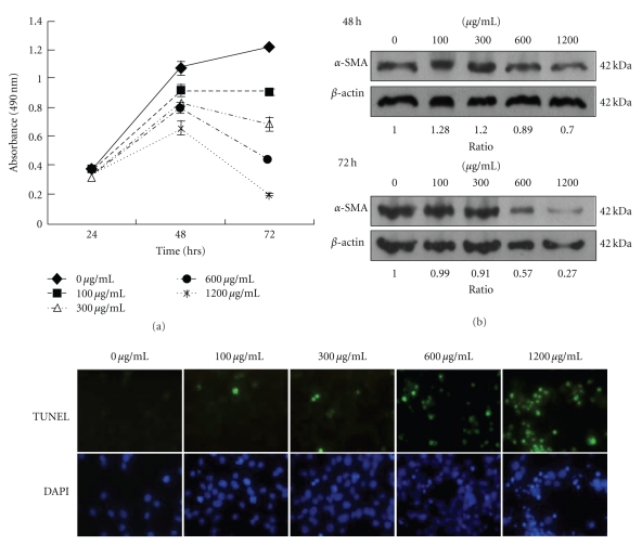
Figure 1.
The mYGJ exhibits potent antifibrosis activity on HSC-T6 cells. (a) HSC-T6 cells were seeded onto 96-well plates and the indicated dose of mYGJ was added to the medium for 24, 48, and 72 hours, respectively. Cell viability was measured by MTS assay as described in the Material and methods. The data represented the mean ± S.D. of three independent experiments. (b) HSC-T6 cells were treated with the indicated concentration of mYGJ. The cell lysates were harvested at 48 and 72 hours posttreatment and were subjected to Western blot analysis using the anti-α-SMA antibody. The expression of β-actin was used for the control of equal protein loading. The band intensity of Bcl-2 or Bax versus β-actin was determined and the relative ratio to control experiment which did not treat with mYGJ was indicated below the data. (c) Representative photographs of HSC-T6 cells that were treated with the indicated concentrations of mYGJ for 72 hours. The determination of apoptosis was performed by TUNEL assay with the apoptotic cells appeared with green fluorescence. Nuclei were counter-stained using DAPI (blue).

