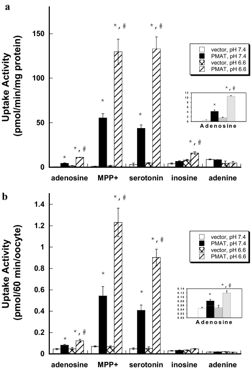
Fig. 4.
a, effect of extracellular pH on PMAT-mediated uptake in MDCK cells. Vector-transfected and PMAT-expressing MDCK cells were incubated with 1 μM concentrations of various 3H-labeled compounds for 1 min at 37°C in the presence of NBMPR (0.5 μM). Each value represents the mean ± S.D. (n = 3). b, effect of extracellular pH on PMAT-mediated uptake in Xenopus oocytes. Water- or PMAT cRNA-injected oocytes were incubated with 1 μM concentrations of various 3H-labeled compounds for 60 min at 25°C in the presence of NBMPR (0.5 μM). Each value represents the mean ± S.E. (n = 8–10). *, p < 0.01, significantly different from vector-transfected cells. #, p < 0.01, significantly different from PMAT activity measured at pH 7.4.

