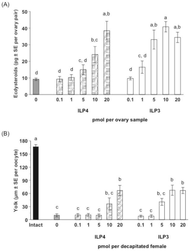
Fig. 2.
ILP4 and ILP3 stimulate ecdysteroid synthesis by ovaries and yolk uptake by primary oocytes from blood-fed, female Ae. aegypti. (A) Ecdysteroid production (pg ± SE) by ovaries treated with ILP4 or ILP3. Ovaries from sugar-fed females (4 ovary pairs per sample) were incubated 6 h in medium containing a given amount of each ILP (0.1–20 pmol), followed by quantification of ecdysteroid amounts in the medium. Ovaries incubated in medium without ILP (0) served as a negative control. Different letters above a given treatment indicates means that significantly differ from one another (F10, 94= 14.6, P<0.0001; followed by comparison for all pairs using the Tukey-Kramer procedure). (B) Yolk uptake (μm ± SE) by primary oocytes following injection of ILP4 or ILP3. Females were decapitated within 2 h after blooding feeding and injected once with a given ILP (0.1–20 pmol). Yolk deposition was then measured 24 h later. Females injected with saline only served as a negative control while normal, nondecapitated (intact) females served as a positive control. A minimum of 12 mosquitoes was analyzed per treatment. Different letters above a given treatment indicates means that significantly differ from one another (F11, 165= 32.9, P<0.0001 and Tukey-Kramer procedure, α=0.05).

