
Figure 8.
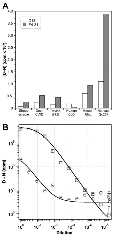
Improved detection of BSE prions. (A) Detection of PrPSc in infected brain homogenates of different species, as indicated, using F4-31 mAb (shaded bars) or HuM-D18 recFab (open bars) as the capture antibody in the CDI. Results are presented as the (D–N) difference in fluorescence signals, measured in counts per minute (cpm), from denatured (D) and nondenatured (N) samples. (B) Measurement of BoPrPSc in BSE-infected bovine brain samples diluted into normal bovine brain homogenate, using either HuM-D18 recFab (circles) or F4-31 mAb (squares) as the capture antibody in the CDI. In both experiments, europium-conjugated HuM-P recFab was used as the detection antibody. The (D–N) difference is directly proportional to the concentration of PrPSc in the sample. Data points and bars represent the averages and standard deviations, respectively, from three independent measurements.