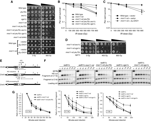
Figure 2.
Phenotype of mre11-nd and mre11-nd sgs1Δ mutants. (A) Suppression of the mre11-nd IR sensitivity by the yku70Δ mutation (quantitation in (B)) or high-copy expression of EXO1 (quantitation in (C), *P=0.03, unpaired t-test). (D) Radiation sensitivity of mre11-nd mutants in conjunction with sgs1Δ or exo1Δ mutations. (E) Schematic representation of the chromosome III MAT locus used in the physical assay to assess resection of an HO-induced DSB. The 5′-3′ degradation destroys the StyI (S) and XbaI (X) recognition sites, which translates into the disappearance of the StyI/XbaI digestion fragments. (F) Southern blot analysis and (G) cut fragment intensity plots showing the kinetics of the cut fragment intensity disappearance as a ratio of the intensity 30 min after induction. The means from four experiments are presented, error bars indicate s.d.

