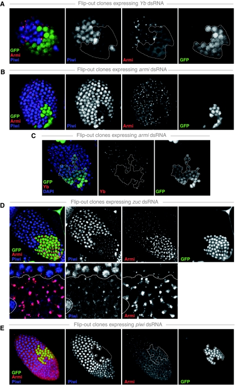
Figure 5.
Comprehensive analysis of Armi, Piwi and Yb localization upon their reciprocal knockdown. (A–E) Shown are surface views of the follicular epithelium of egg chambers containing clones expressing dsRNA hairpins against the indicated genes stained for Piwi (blue), Armi (red) and Yb (red). Clones are marked by the presence of GFP (green) and clone borders are indicated (dashed line). In all rows, the merged RGB image is shown alongside with the individual channels in black and white. In (D), the lower row shows higher magnification views.

