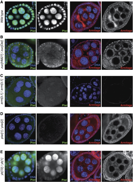
Figure 8.
Similarities of Piwi biology in soma and germline of the ovary. (A–E) Shown are optical sections through mid stage egg chambers of indicated genotypes (left) stained for DNA (blue), Piwi (green) and Armi (red). Peri-nuclear accumulation of Piwi in germline cells of zuc mutant ovaries is marked by asterisks in (B).

