Fig. 1.
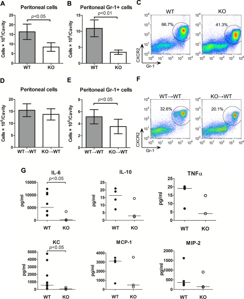
Knockout (KO) and KO→wild-type (KO→WT) mice have impaired neutrophil mobility and reduced peritoneal cytokine/chemokine levels. Mice were injected intraperitoneally with thioglycollate. Ten hours after the injection, 3 ml of normal saline was injected and mixed well. The peritoneal lavage fluid and leukocytes were isolated and analyzed. A: total numbers of cells recruited into the peritoneal cavity of WT and KO mice. B: total neutrophils recruited into the peritoneal cavity of WT and KO mice. Each error bar represents mean ± SD of 3 mice. C: a representative example of flow cytometry plots of peritoneal neutrophils from WT and myeloid differentiation factor 88 (MyD88)-deficient mice (MyD88−/−). The cells were gated on CXCR2 and Gr-1. The cells within the circle represent CXCR2+/Gr-1+ neutrophils, and the percentage of neutrophils is shown. D: total numbers of cells recruited into the peritoneal cavity of the chimeric mice. E: total neutrophils recruited into the peritoneal cavity of the chimeric mice. Each error bar represents mean ± SD of 3 mice. F: a representative example of flow cytometry plots of peritoneal neutrophils from WT→WT and KO→WT chimeric mice. G: peritoneal lavage was analyzed for cytokine production using Luminex multiplex immunoassay. The bars in each dot plot represent median values of the measured cytokines. Some cytokine values were overlapping. n = 4–7. MCP-1, monocyte chemoattractant protein 1; MIP-2, macrophage inflammatory protein 2.

