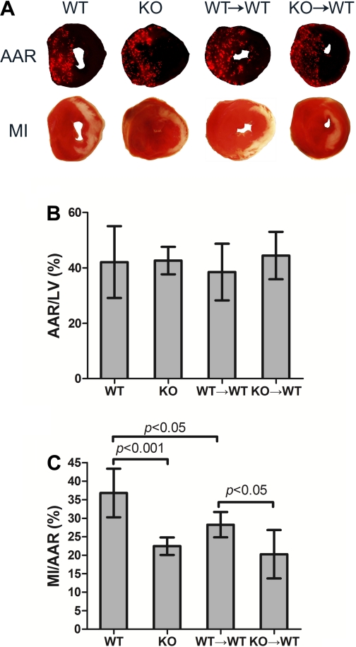
Fig. 4.
KO→WT mice have smaller myocardial infarction (MI) size compared with WT→WT mice. Mice were subjected to 30 min of ischemia and 24 h of reperfusion. At the end of reperfusion, animals were euthanized, and area-at-risk (AAR) and MI were analyzed. A: representative pictures of AAR and MI from the 4 groups of mice. B: cumulative data of AAR/left ventricle (LV). C: cumulative data of MI/AAR. Each error bar represents mean ± SD of 6–9 mice.

