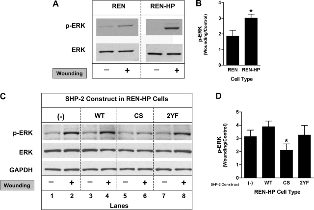
Fig. 6.
Effects of the expression of SHP-2 constructs on wound-induced ERK activation in REN and REN-HP cells. Cell lysates from nonwounded or wounded monolayers of REN or REN-HP cells were immunoblotted with anti-phopho-ERK and anti-ERK antibodies. A: expression of PECAM-1 in REN cells resulted in increased phospho-ERK levels after wounding compared with nontransfected REN cells. B: densitometry confirmed that ERK activation following wounding of REN cells (expressed as the ratio of wounding/control) was increased by the expression of PECAM-1. Data are presented as means ± SE (n = 4; *P < 0.05, compared with REN cells). C: expression of the CS-SHP-2 (CS) construct in REN-HP cells inhibited ERK activation (lanes 5 and 6 compared with lanes 1 and 2) following wounding, while the presence of WT-SHP-2 (WT) or 2 YF-SHP-2 (2YF) did not suppress PECAM-1-dependent, wound-induced, ERK activation (lanes 3 and 4, and lanes 7 and 8, compared with lanes 1 and 2). D: densitometry confirmed that the ERK activation mediated by PECAM-1 (expressed as the ratio of wounding/control) was suppressed by coexpression of CS-SHP-2. Data are presented as means ± SE (n = 3; *P < 0.05, compared with REN-HP/WT cells). The densitometry were all initially normalized to total ERK expression. (The data presented in A and C are from the same experiment and the same gel for each antibody. They are representative of 3–4 experiments).

