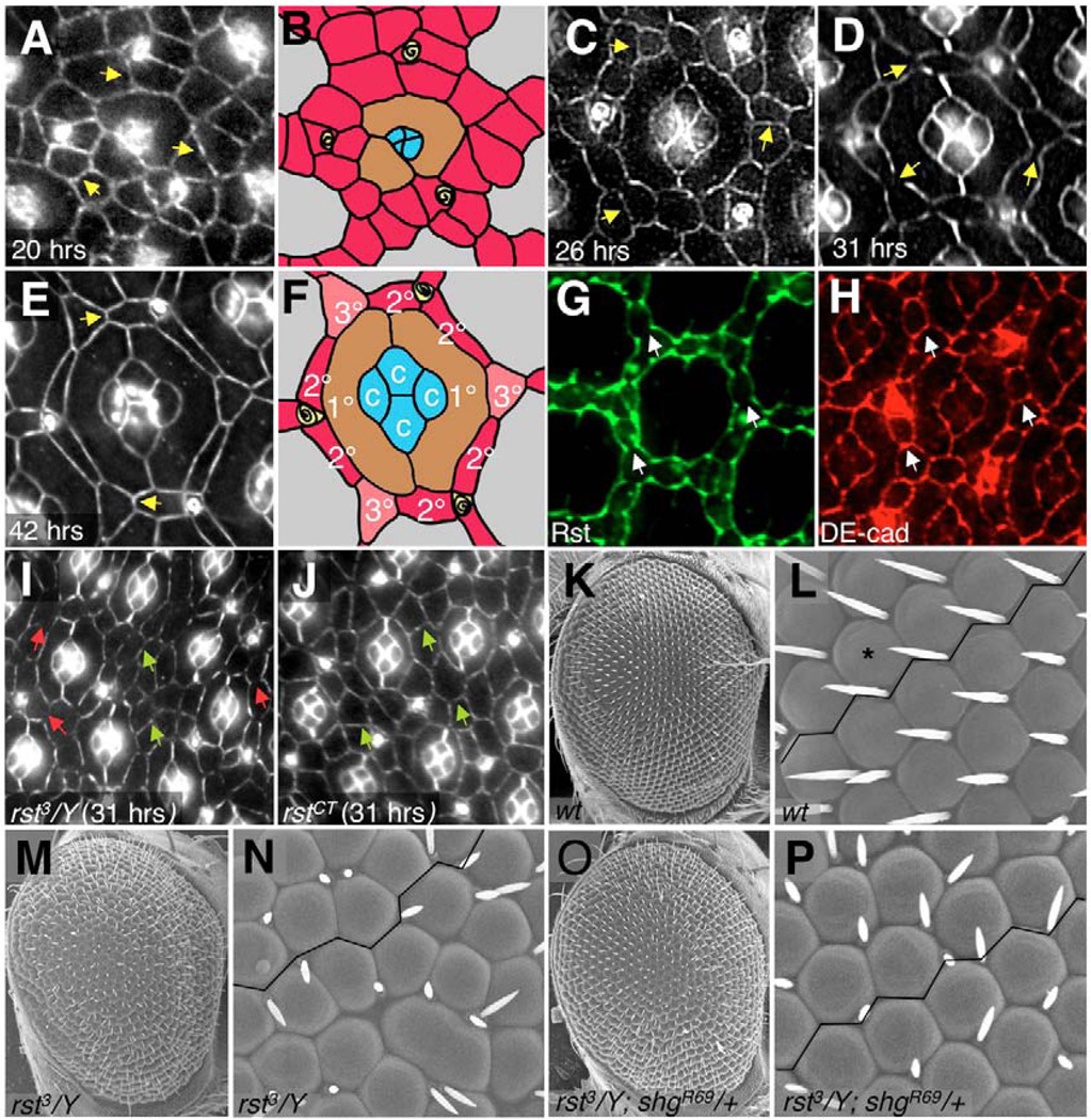
Fig. 1. Morphogenesis of the Drosophila pupal retina and the role of cell adhesion.
(A–F) Time course of retinal development. Apical cell profiles were visualized with anti-Armadillo to highlight adherens junctions. Anterior is to the right; times refers to hours after puparium formation (APF). Unpatterned arrays of IPCs (A,B) sort to single file (C); IPC number continues to decrease as patterning tightens (D) until the final pattern is achieved (E,F). B and F are schematics of the central ommatidium from A and E, respectively; cone cells (c, blue shading), primary pigment cells (1°, brown), IPCs (red), 2° (red), 3° (pink) and bristles (yellow) are indicated. Arrows in A,C,D,E point to IPC:IPC adherens junctions. (G,H) 26 hours APF retina stained with anti-Rst (G) and anti-DE-cadherin (H). Arrows point to the IPC:IPC junctions, where DE-cadherin is expressed and Rst is absent. (I,J) 31 hours APF rst mutant retinas; magnification is reduced to show additional ommatidia. Green arrows point to IPC:IPC junctions that failed to clear (compare with wild type, D). This defect correlated with the failure in mutant IPCs to sort out into single-cell rows, as observed in mutants for the hypomorphic allele rst3, which is subject to position effect variegation (I). Red arrows in I point to rst3 regions where IPCs have sorted out into single layers and have also cleared out their junctions; these are likely to contain normal levels of Rst protein. (K–P) Scanning electron micrographs of adult eyes (genotypes as listed) taken at 180× (K,M,O) and 800× (L,N,P). K and L show a wild-type adult eye; a single ommatidium is indicated with an asterisk. Note the straight ommatidial rows, highlighted by a line drawn between ommatidia. The aberrant ommatidial packing observed in an rst3 eye (M,N) is rescued by removing a single functional copy of shg (O,P).

