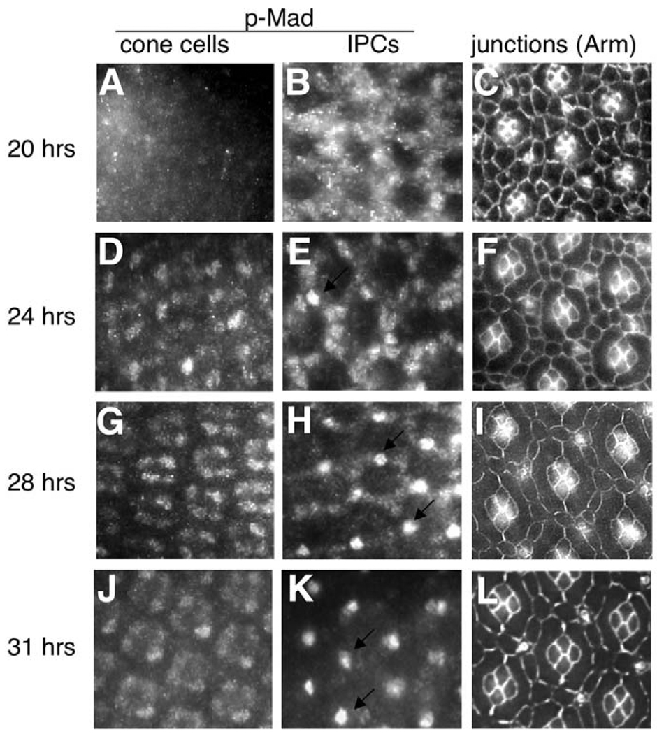
Fig. 4. Dynamic Dpp signaling activity in the Drosophila pupal retina.
(A–L) Dpp pathway activity is visualized in pupal retinas at different developmental stages using anti-p-Mad antibody. (A,D,G,J) p-Mad staining at the level of the cone cell nuclei; (B,E,H,K) p-Mad staining at the level of the IPC nuclei. (C,F,I,L) The maturing IPC pattern from age-matched retinas as development proceeded; cell membranes were stained with anti-Armadillo antibody. Time refers to hours APF. Arrows in E,H,K point to nuclear p-Mad in the sensory bristles.

