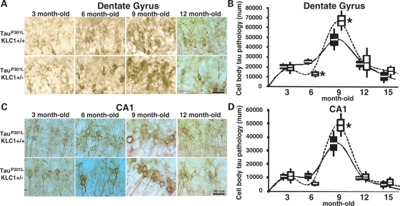
Figure 2.
Increased cell body pathology in brains of KLC1 reduction combined with tau P301L. (A and C) Staining of phosphorylated tau using CP13 antibody. Neuronal cell bodies with abnormal p-tau accumulation are shown for dentate gyrus (A) and CA1 (C) region of the hippocampus in 3-, 6-, 9- and 12-month-old tauP301L;KLC1+/+ and tauP301L;KLC1+/− mice. (B and D) Stereological quantification of neuron numbers showing p-tau cell body accumulation in dentate (B) and CA1 (D) hippocampus for tauP301L;KLC1+/+ (black) and tauP301L;KLC1+/− (27). Distribution of the average number is plotted. Boxes and bars correspond to SEM and STDEV, respectively. Mann–Whitney, non-parametric test; n = 4, *P < 0.05.

