Figure 1.
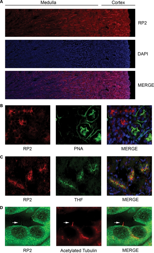
RP2 is enriched in distinct regions of the kidney. (A) RP2 staining in mouse kidney. PFA-perfused frozen mouse kidney sections were permeabilized and stained with anti-RP2 antibodies (red) and DAPI (blue) to mark nuclei. (B) RP2 does not localize to distal convoluted tubules. Mouse kidney sections were stained as above with the addition of peanut lectin agglutinin (PNA-FITC) to label distal convoluted tubules. (C) RP2 localizes to Tamm Horsfall protein (THF) positive (green) segments of the nephron. Mouse kidney sections were stained as above with the addition of anti-THF antibody. (D) RP2 localizes to primary cilia in mouse kidney tubules. Mouse kidney sections were stained as above with the addition of anti-acetylated tubulin to label primary cilia (red).

