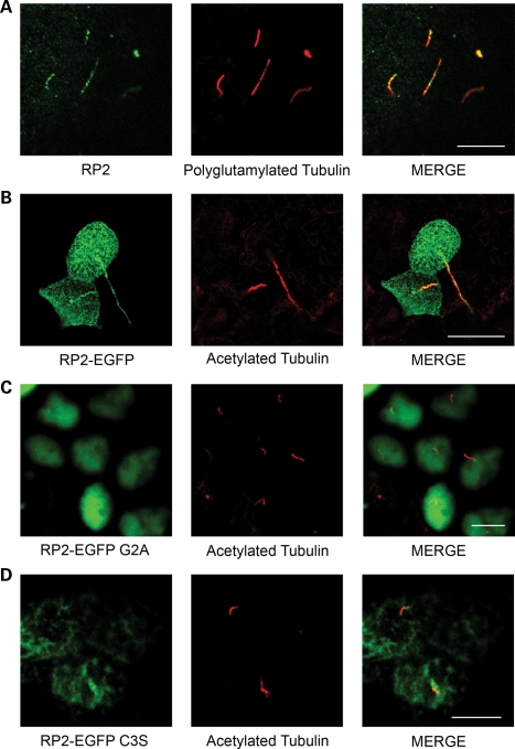
Figure 2.
RP2 localizes to primary cilia. (A) RP2 localizes to cilia in MDCK cells. MDCK cells were grown 7 days post-confluence, fixed, permeabilized and immuno-stained using antibodies against RP2 (green) and polyglutamylated tubulin (red). (B) RP2-EGFP localizes to primary cilia. MDCK cells stably expressing EGFP-tagged RP2 (green) were grown for 7 days post-calcium switch to allow cilia formation. Cilia were visualized using an antibody against the cilia marker acetylated tubulin (red). (C) Mutation of the N-terminal myristoylation motif prevents cilia localization of RP2-EGFP and mistargets it to nuclei. MDCK cells stably expressing RP2-EGFP G2A (green) were grown for 7 days post-confluence on trans-well filters, fixed, permeabilized and immuno-stained with an anti-acetylated tubulin antibody (red). (D) RP2-EGFP C3S fails to target to cilia. MDCK cells stably expressing the palmitoylation mutant C3S (green) were treated as above. All scale bars (white) represent 10 µm.

