Figure 3.
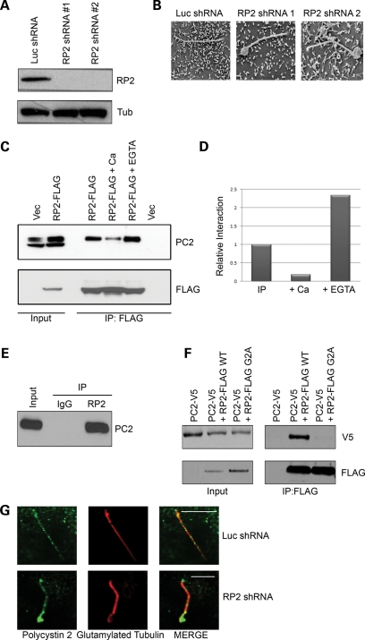
RP2 forms a complex with polycystin 2. (A) Knockdown of RP2 by retroviral shRNA. MDCK cells were retrovirally infected with either a luciferase-specific shRNA construct (Luc) or one of two separate canine-specific RP2 shRNA constructs (RP2 shRNA no. 1 and RP2 shRNA no. 2) and stable pools selected. Cell lysates were subjected to SDS–PAGE and western blotting with the indicated antibodies. (B) Ablation of RP2 results in swelling of the cilia tip as shown by SEM. Control or RP2 shRNA were grown 7 days post-confluence on trans-well filters, fixed and prepared for SEM imaging. (C) Exogenous RP2 co-immunoprecipitates endogenous polycystin 2. Cell lysates (Input) from MDCK cells stably expressing either empty vector (pQCXIP) or RP2-FLAG wild-type (WT) were subjected to anti-FLAG immunoprecipitation in the presence or absence of 5 mm calcium chloride or EGTA. Immunoprecipitates were washed and subjected to SDS–PAGE and western blotting with the antibodies indicated. (D) Quantification of the effect of calcium and EGTA on the co-immunoprecipitation of RP2 and polycystin 2. The western blots in (C) were subjected to densitometric analysis. (E) Endogenous RP2 forms a complex with endogenous polycystin 2 in retina. Lysate (Input) from bovine retina was subjected to anti-RP2 immunoprecipitation. Immunoprecipitates were washed and subjected to SDS–PAGE and western blotting with the antibodies indicated. (F) The membrane association of RP2 is required for the interaction with polycystin 2. HEK293 cells were transiently transfected with polycystin 2-V5 and either wild-type (WT) RP2-FLAG or the membrane targeting mutant (G2A) RP2-FLAG. Lysates were subjected to immunoprecipitation as described above. (G) Ablation of RP2 results in accumulation of endogenous polycystin 2 at the distal region of cilia. Control (Luc shRNA) or RP2 shRNA were grown 7 days post-confluence on trans-well filters, fixed and stained with antibodies against polycystin 2 (green) and glutamylated tubulin (red). All scale bars (white) represent 10 µm.

