Figure 5.
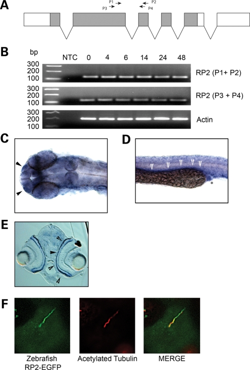
RP2 expression in zebrafish. (A) Gene structure of the Zebrafish rp2. Protein coding region shown in grey. Shown are the primer sets used for RT–PCR. (B) RT–PCR analysis of rp2 transcript at different time points post-fertilization (hpf). NTC, no template control. (C) rp2 is expressed in the olfactory placode (arrowheads). In situ hybridization was performed on 24 hpf embryo with an rp2 antisense probe. (D) rp2 is expressed in the pronephric duct at 24 hpf. In situ hybridization was performed on a 24 hpf embryo with an rp2 antisense probe. Arrowheads show signal along the pronephric duct and asterisk shows cloaca. (E) rp2 is expressed in the photoreceptor layer (arrowheads) of the retina at 72 hpf. In situ hybridization was performed on a 72 hpf embryo with an rp2 antisense probe. (F) Zebrafish RP2 localizes to cilia in MDCK cells. MDCKII cells stably expressing zebrafish RP2-EGFP were grown for 7 days post-calcium switch to allow cilia formation. Cilia were visualized using an antibody against the cilia marker acetylated tubulin (red).

