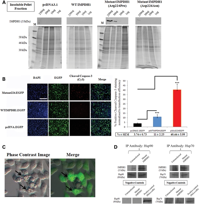
Figure 3.
Formation of insoluble protein aggregates and induction of cell death by mutant IMPDH1 in HeLa cells. (A) Cellular fractionation assay illustrating dose-dependent insoluble mutant IMPDH1 protein aggregate formation in HeLa cells. Note that mutant IMPDH1 bearing the Arg224Pro mutation accumulated in the insoluble protein fraction, giving off a strong signal at 250 and 50 ng. In contrast, no or weak IMPDH1 was detected in HeLa cells transfected with empty vector, wild-type IMPDH1 and mutant IMPDH1 bearing the Asp226Asn mutation. Coomassie blue staining of identical gels as loading controls. M, protein marker. (B) Representative fluorescent images of cleaved caspase-3 staining in HeLa cells transfected with WT or mutant IMPDH1 EGFP fusion expression plasmids. Bar charts represent percentage positive cleaved caspase-3 staining normalized against transfection efficiency (***P < 0.0001). Scale bar denotes 200 μm. Data presented here represent the mean of three independent experiments carried out in triplicate. Error bars denote standard error of the mean (SEM). (C) Identification of cell blebbing (indicated by arrows) in HeLa cells transfected with mutant IMPDH1 bearing the Arg224Pro mutation. Merge = Phase contrast image overlaid with EGFP. Scale bar denotes 40 μm. (D) Hsp70 and Hsp90 immunoprecipitate with mutant IMPDH1: western blot analysis in HeLa cells after immunoprecipitation with IMPDH1 antibody, with total cell lysates as positive controls and isotype-matched IgG as negative controls. Immunoprecipitation antibodies as loading controls.

