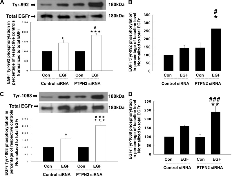
Fig. 2.
Loss of PTPN2 enhances phosphorylation of the EGFr tyrosine residue Tyr-992 and Tyr-1068 in response to EGF. Either control siRNA- or PTPN2 siRNA-transfected T84 cells were treated with EGF (100 ng/ml) for 5 min. Analyses were performed using whole cell lysates. A: representative Western blots show phosphorylation of the EGFr residue Tyr-992 and expression of total EGFr. Below is the densitometric analysis of 3 similar experiments. Data are presented as percentage of the respective controls. B: secondary densitometric analysis demonstrates the magnitude of Tyr-992 induction from baseline level (analysis was performed as described in Fig. 1C) in percentage of untreated cells transfected with control siRNA. C: phosphorylation of the EGFr residue Tyr-1068 in response to EGF and expression of total EGFr is demonstrated by representative Western blots. The densitometric analysis of 5 similar experiments is shown in the histogram below. Data are presented as percentage of the respective controls. D: secondary densitometric analysis demonstrates the magnitude of Tyr-1068 induction from baseline level (analysis was performed as described in Fig. 1C) in percentage of untreated cells transfected with control siRNA. Significant difference vs. the respective control: *P < 0.05, **P < 0.01, ***P < 0.001. #P < 0.05, ###P < 0.001 vs. EGF treatment of T84 cells transfected with control siRNA.

