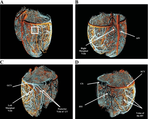
Fig. 1.
Rendering of the reconstructed arterial and venous trees (orders 1 to 11 and −1 to −12) as viewed form four different aspects: anterolateral left (A), anterolateral right (B), posterolateral left (C), and posterolateral right (D). The rendering was done using POVWIN Raytracer. White rectangle in A marks the location chosen for the close-up image in Fig. 2, A–D. Orange, arterial; cyan, venous. AIV, anterior interventricular vein; GCV, great cardiac vein; LV, left ventricle; CS, coronary sinus; PIV, posterior interventricular vein; SCV, small cardiac vein; RV, right ventricle.

