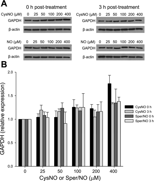
Fig. 2.
GAPDH protein levels. BAECs were incubated for 1 h with CysNO or Sper/NO in HBSS. Cells were washed and either harvested immediately (0 h posttreatment) or incubated further for 3 h in 2% FBS medium (3 h posttreatment). GAPDH levels were assessed by Western blot analysis. A: representative Western blot of GAPDH. NO, nitric oxide, represents the concentration of Sper/NO after nitric oxide. B: densitometry analysis of GAPDH levels normalized to β-actin. Data represent means ± SE (n = 3 experiments).

