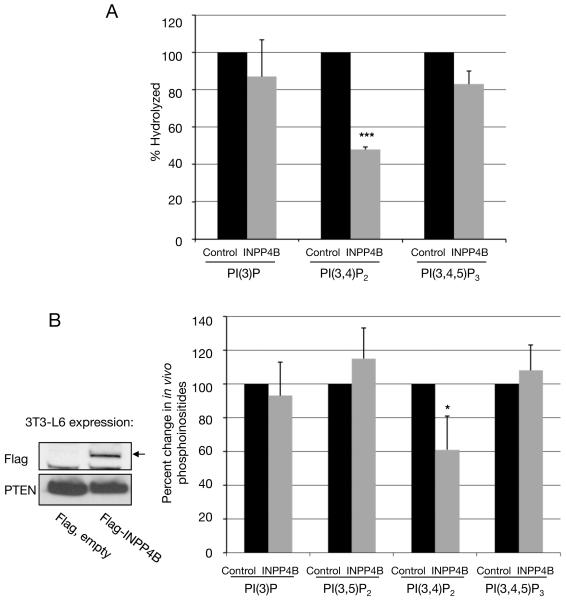
Figure 5. Substrate specificity of INPP4B.
(A) FLAG-tagged INPP4B was immunoprecipitated from 293T cells and incubated with either [32P]-PI(3)P, or [32P]-PI(3,4)P2 or [32P]-PI(3,4,5)P3. As control, the phosphoinositides were incubated with Flag-immunoprecipitate from transfected 293T cells expressing an empty Flag-expression construct. The percent hydrolysis of each lipid was determined by chloroform/methanol/HCL extraction, thin layer chromatography and Phosphoimager analysis. (n=3: ***p < 0.001). (B) Overexpression of human INPP4B causes a reduction in cellular PI(3,4)P2 levels in vivo. 3T3-L6 cells were transiently transfected with empty Flag or Flag-INPP4B constructs and labeled with [32P]-inorganic phosphate. Lipids were extracted, deacylated and the headgroups separated by HPLC. The radioactivity in the glyceroyl-phosphoryl-inositol moieties of each of the D-3 phosphorylated phosphoinositides was then normalized to the total radioactivity in the more abundant phosphoinositides, PI(4)P and PI(4,5)P2 (which did not significantly vary between experiments). The bars indicate per cent changes in these ratios in INPP4B transfected cells compared to control cells. The actual ratios of radioactivity in each lipid to total radioactivity in PI(4)P plus PI(4,5)P2 in the control 3T3-L6 cells were: PI(3)P – 3.2%, PI(3,5)P2 – 0.035%, PI(3,4)P2 – 0.17%, PI(3,4,5)P3 – 0.69%. (Averages from three experiments are presented: *p<0.1). Protein expression of Flag-INPP4B in 3T3-L6 cells was demonstrated by Immunoblotting. Protein loading was shown in using antibody directed against PTEN. (Inset). All data are shown as mean +/− SD.

