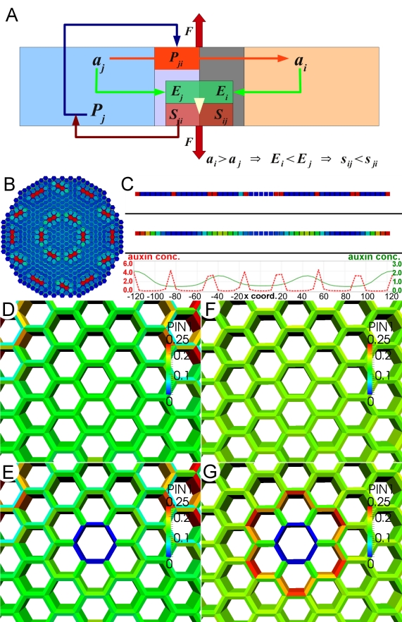
Figure 7. Mathematical model of auxin transport and mechanical stress.
(A) Schematic representation of the interactions leading to a pattern-forming behavior in the model. Auxin (a) is transported out of cell j to cell i by the PIN1 (P) proteins localized to the membrane, Pji. Auxin concentration in each cell affects the elasticity (Ei and Ej) of the adjacent wall, which influences the mechanical stress (Sij and Sji) perceived in both parts of the wall between the cells as a result of the force F. For example ai>aj leads to Ei<Ej, which in turn causes Sij<Sji. The cycling of PIN1 between cytosol, Pj, and the membrane, Pji, depends on these stresses, and larger Sij causes stronger allocation of Pj to Pji. (B) Example of the auxin pattern created spontaneously in the model by applying uniform tension to a two-dimensional template. (C) Spacing of emerging auxin peaks can be controlled by adjustment of model parameters. Patterns of auxin distribution obtained in two different one-dimensional simulations of the model show different arrangements of auxin peaks (compare red versus green peak profiles). (D–G) Comparison of the behavior of the new model with a previously proposed auxin transport model [1] in the case of the response to ablation of a cell. In the auxin-concentration-based model, removal of the cell in the region of uniform auxin distribution (D) causes only minor response of the PIN1 polarization in the cells neighboring the removed cell (E). In the stress-based model, we observed much stronger, outward PIN1 polarization in the cells closest to the ablated region (G), as compared to the situation prior to ablation (F). The ablation results for the new model provide a better fit to the experimental data (see Figures 2 and 4).

