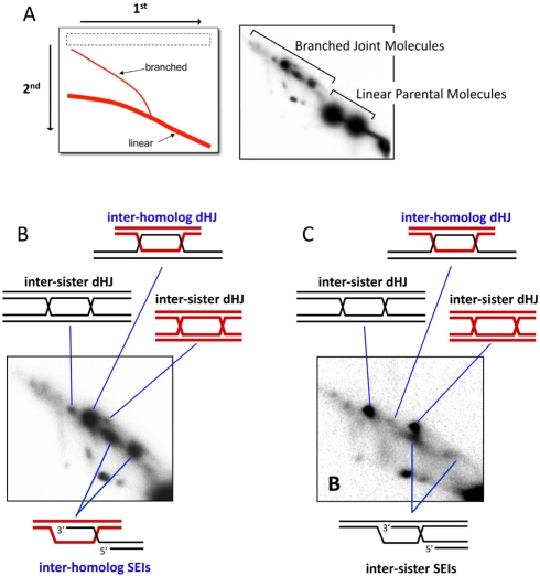
Figure 3. Monitoring template choice by 2-D gel electrophoresis.
(A) The second dimension of a 2-D gel accentuates the shape element of DNA molecules such that branched species migrate more slowly than linear duplexes of identical mass. The right hand panel shows detection of JM intermediates via Southern hybridization of a 2-D gel. The analyzed locus contains restriction-site polymorphisms between the two parental homologs. (B) Close-up of the JMs in (A), highlighting the SEI and dHJ intermediates. Note the preponderance of inter-homolog dHJs relative to the inter-sister dHJs. (C) 2-D gel analysis of a mutant with a defect in template choice. In this strain, inter-homolog dHJs are almost absent and nearly all JMs, both SEIs and dHJs, are formed between sister chromatids.

