Fig. 1.
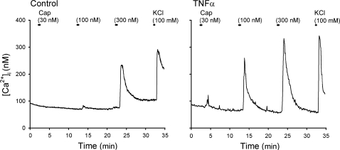
Representative experimental records illustrating the change in intracellular Ca2+ concentration ([Ca2+]i) evoked by increasing concentrations of capsaicin (Cap) in vagal sensory neurons labeled with 1,1′-dioctadecyl-3,3,3′,3′-tetramethylindocarbocyanine perchlorate (DiI). Left: a control neuron (diameter 33.3 μm) incubated with vehicle (culture medium: modified DMEM/F-12 solution) for ∼24 h. Right: a neuron (diameter 24.8 μm) incubated with TNFα (50 ng/ml in culture medium) for ∼24 h. Both neurons were isolated from nodose ganglia of the same rat (185 g). Cap was applied for 30 s each, and KCl solution (100 mM, 15 s) was applied to test cell vitality at the end of both experiments.

