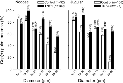
Fig. 3.
Effect of TNFα pretreatment on the size distributions of Cap-sensitive pulmonary sensory neurons. Cells were grouped based on their size (in 5-μm increments of cell diameter), and Cap(+) neurons were depicted as the percentage of all the DiI-labeled nodose and jugular ganglion neurons that were activated by KCl in each size category. A neuron was considered Cap(+) if its Cap (300 nM)-evoked Ca2+ transient exceeded 20% of its peak Ca2+ transient generated by KCl (100 mM). Closed and open bars represent treated neurons (pretreated with 50 ng/ml TNFα for ∼24 h) and control neurons (pretreated with vehicle for ∼24 h), respectively. To have a sufficient number of cells for a more accurate analysis, the numbers of small (<20 μm) and large (>35 μm) cells higher than their normal distribution were selected and tested in this study (see text for details).

