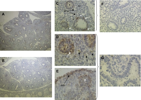
Fig. 3.
mTORC1 activity in the pseudoglandular rat lung in vivo. Gestation day 19 fetal rat lungs were fixed and stained with a phospho-antibody against the mTORC1 substrate, p70S6K1-T389. A: low-power (×40) image of fetal lung in situ. p70S6K-T389 phosphorylation is apparent in the nascent airway epithelium (brown stain). B: control section treated with nonimmune IgG at same magnification. C: mid-power (×200) image showing airway (a), mesenchyme (m), and endothelial tube (et) structures. Note apparent polarization of mTORC1 activity around the tube (white arrows). D: high-power (×400) image showing mTOR activity extending laterally from the epithelium indicating activity within a possible branching node. Localized activity in cells of the mesenchyme is evident (black arrows). E: high mTOR activity in the mesothelial lung lining (mth), a site of FGF9 expression (56). White arrow indicates regional activity in an epithelial lateral branch node. F and G: ×200 and ×400 images of sections treated with nonimmune IgG.

