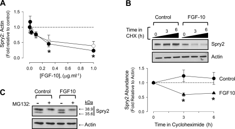
Fig. 8.
FGF-10 destabilizes Spry2 in FDLE. A: abundance of Spry2 declines with increasing FGF-10 concentration at fetal (closed circles) and alveolar (open circles) Po2. Densitometry performed on full-length Spry2 in ratio to actin. *P < 0.05 relative to control, n = 4. B: Spry2 becomes unstable in the presence of FGF-10. FDLE were treated with 100 μM cycloheximide (CHX) and 0.1 μg·ml−1 FGF-10 as indicated at fetal Po2. Representative blot is accompanied by graph showing full-length Spry2 abundance measured in ratio to actin. Circles, control; triangles, 0.1 μg·ml−1 FGF-10. *P < 0.05, n = 4 relative to time 0. Error bars may be within symbols. C: Spry2 cleavage is proteasomal. FDLE were treated with 10 μM MG132 and 0.1 μg·ml−1 FGF-10 at fetal Po2 for 3 h as indicated. Proteins were separated by 10% SDS-PAGE to resolve both full-length and cleaved Spry2 product. Blot is representative of n = 3.

