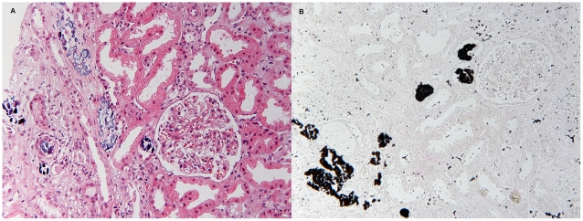
Figure 1. Renal biopsy findings in acute phosphate nephropathy.
A) Abundant calcifications are seen within tubules and in the interstitium. Adjacent tubules are athrophic and there is interstitial fibrosis (Hematoxylin and eosin staining, original magnification x 400). B) Positive von Kossa staining in the same biopsy confirms that the calcifications are composed of calcium phosphate (original magnification x 400).

