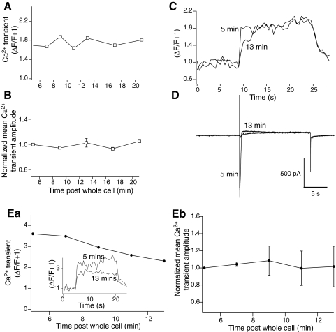
Fig. 7.
Inhibitor 2 did not cause a time-dependent modification of Ca2+ transients recorded following step protocols with either no synaptic or no NMDA component. Neurons were recorded as for Figs. 5 and 6 but no stimulation of synaptic inputs to the CA1 region was applied. A: peak amplitude of stimulus evoked fluorescence transients obtained during a 20 s voltage step to –40 mV, measured in the distal dendrites for time points from 5 to 21 min after obtaining whole cell access. B: normalized peak amplitude of evoked fluorescence transients over time after whole cell access in all controls for all neurons recorded. C: traces from cell in A showing fluorescence transients comparing responses from stimuli 5 min post whole cell access to 13 min after obtaining access from distal dendrites. D: current traces recorded at 5 and 13 min after obtaining whole cell access. These were recorded simultaneously to the fluorescence transients shown in C with the same time bases. Ea: peak amplitude of stimulus evoked fluorescence transients recorded in d-AP5 (50 μM) obtained during a 20 s voltage step to –40 mV while stimulating at 2 s intervals (as in Fig. 5) measured in the distal dendrites for time points from 5 to 13 min after obtaining whole cell access. (Inset: examples of Ca2+ transients at 5 and 13 min after obtaining whole cell access.) Eb: normalized peak amplitude of evoked fluorescence transients over time after whole cell access all neurons recorded under conditions in Ea.

