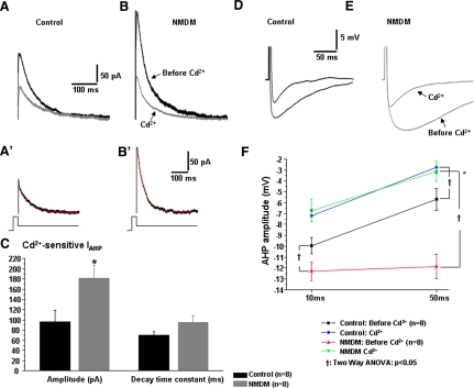
Fig. 9.
Dependency of maternal diabetes-induced increase of AHP currents (IAHP) and AHP on Ca2+influx in PCMNs. The IAHP current and AHP were evoked and measured as in Fig. 8. In addition, AHP was evoked by a current injection of sufficient intensity to generate a single AP. The peak of AHP was measured at 10 and 50 ms. A: (control) and B (NMDM). The black trace is the IAHP before Cd2+, and the gray trace is the IAHP after Cd2+ application. A′ (control) and B′ (NMDM). The Cd2+–sensitive IAHP was obtained by subtracting the current after Cd2+ from the current before Cd2+ application. The decay of Cd2+–sensitive IAHP was fitted by a single exponential equation (red line). C: maternal diabetes significantly increased Cd2+–sensitive IAHP (*P < 0.05) but did not increase decay time constant. D and E: representative AHP waveforms of control (D) and NMDM (E) PCMNs before and after Cd2+ application. F: Cd2+ significantly reduced AHP in both groups (†P < 0.05). After application of Cd2+, the AHP amplitude at 10 and 50 ms in NMDM was comparable to control, indicating that blockade of Ca2+influx completely abolished maternal diabetes-induced increase of AHP. In addition, maternal diabetes induced a significant greater reduction of AHP compared with control (*P < 0.05).

