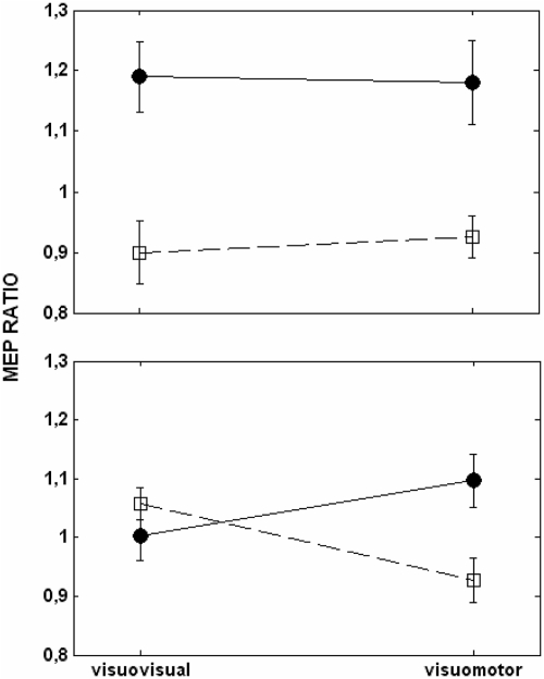
Fig. 3.
Corticospinal excitability of 1st dorsal interoseus (FDI). Motor evoked potentials were measured for the FDI during the maximal aperture of finger movements (top) and during the presentation of the cue (bottom) after visuovisual and visuomotor training. Shown are the means ± SE of the ratio obtained from adjusting the MEPs for the index (●) and the little finger (□) by the baseline.

