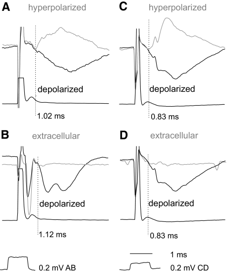
Fig. 3.
Onset of IPSPs evoked from motor nuclei defined by comparing intracellular records in depolarized and hyperpolarized neurons as well as intracellular and extracellular records. Records from 3 VSCT neurons (A, B, and C, D) and cord dorsum potentials. Black traces: during depolarization by 10–15 nA. Gray traces in A and C, during hyperpolarization by 5 and 15 nA and the ensuing reversal of the IPSPs; gray traces in B and D, extracellular records. The pairs of traces in each panel were superimposed so that both stimulus artifacts and the traces directly preceding the IPSPs overlapped. The points of deviation between them are indicated by the vertical dotted lines and show the onset of the IPSPs otherwise not sharp enough to be estimated.

