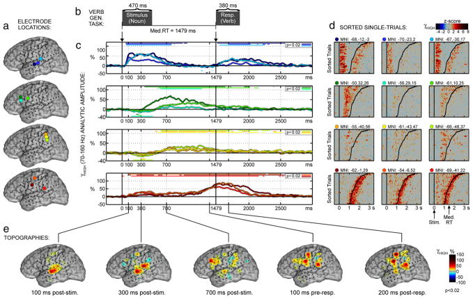
Fig. 3.
Verb generation results for Patient 2. Plotting conventions same as Fig. 2. The second electrode group (green colors) includes sites on the inferior and middle frontal gyri (IFG, MFG). The grid placement was more posterior in this patient, so no coverage was obtained anterior to pars opercularis of the IFG, but coverage of the inferior parietal lobe (IPL) was gained. The third electrode group includes three IPL electrodes on the posterior SMG. The fourth group (red colors) includes one electrode from the TPJ cortex just posterior to the branch point of the Sylvian fissure. Although it is a surprising location from which to observe response-related activity, this electrode responded similarly to the two premotor electrodes and is therefore grouped with them.

