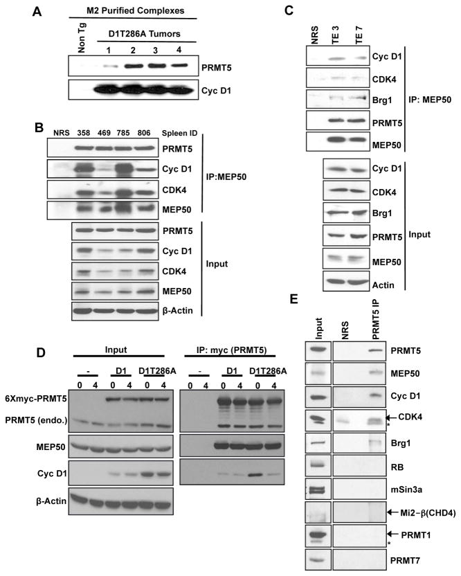
Figure 1. Identification of cyclin D1T286A/PRMT5/MEP50 complexes.
(A) M2 (anti-Flag) purified complexes from Eμ-D1T286A (Flag epitope) transgenic tumors and non-transgenic spleens were blotted for PRMT5 and cyclin D1. (B) MEP50 immunoprecipitates (IP, 1mg whole cell lysate input) prepared from Eμ-D1T286A transgenic tumors were immunoblotted as indicated (C) MEP50 IP (1.5mg whole cell lysate input) from human esophageal cancer cell lines TE3, TE7 were immunoblotted as indicated (Top panel). Input lysates have been shown in the bottom panel. (D) HeLa cells transfected with either wild type cyclin D1 or D1T286A along with myc-PRMT5 and CDK4 were synchronized with aphidicolin. Cells were collected at 0 hours (G1/S boundary) and 4 hours (S-phase) after release. Myc-PRMT5 was pulled down using myc antibody (1mg whole cell lysate input) and immunoblotted as indicated (right panel), input lysates (left panel). (E) Cyclin D1/CDK4 complexes were isolated from Eμ-D1T286A lymphomas using M2-agarose followed by elution with Flag peptide. Eluted complexes were immunoprecipitated with normal rabbit serum (NRS) or PRMT5 antibody. 300μg of eluted complex served as input in NRS or PRMT5 IP, and western analysis was performed as indicated. See also Figure S1 and Table S1.

