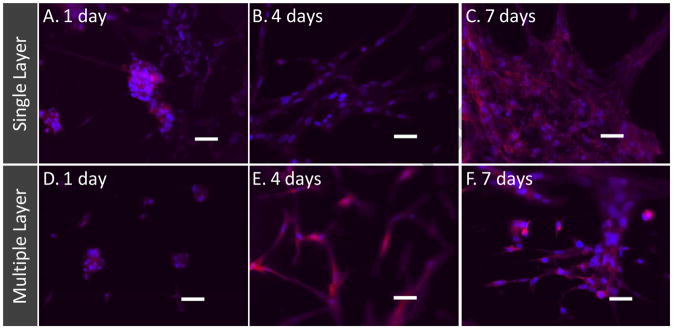
Figure 5. Cell colonization in thin layers using layer-by-layer assembly.
(A–C) Fluorescent micrographs of cells on single layer of fibers. (D–F) Fluorescent micrographs of cells on three layers of fibers. The samples were stained with Alexa phalloidin for cytoskeletal actin (red) and counterstained with DAPI for nuclei (blue) of cells. Scale bar corresponds to 50 μm.

