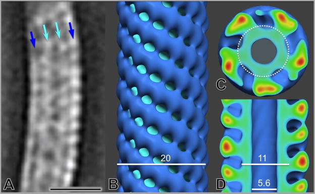
Figure 7.
Structural characterization of periplasmic flagella in situ. A single slice from the averaged map of periplasmic flagella (thick filament) segments illustrates the curved configuration of their superhelical conformation (A). After mathematical ‘straightening’ of the superhelical curvature, the refined structural model of the periplasmic flagella in the surface view (B) and center sections (C and D) reveals the central channel, the filament core and its surrounding protein sheath. The scale bar is 20 nm.

