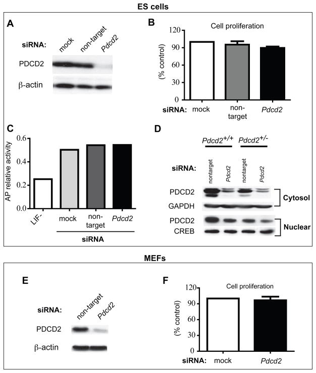
Figure 4. Small amounts of PDCD2 are sufficient for cell survival.
siRNA knockdown of Pdcd2 in ESCs and immortalized MEFs was performed and monitored by various metrics. The knockdown efficiency was assessed in ESCs and MEFs by Western blot analysis (A, E). 72 hours after siRNA transfection, the cells were harvested and counted for proliferation assays (B, F). The cell number was quantified from three independent experiments and shown as the mean ± SEM (% of control). (C) Effect of decreased PDCD2 on ESC pluoropotency was assessed by alkaline phosphatase (AP) activity. The LIF withdrawal group is a positive control for cell differentiation. (D) Western blot analysis of cytosolic and nuclear PDCD2 in Pdcd2 knockdown ESCs. Blots were probed with antibodies indicated. GAPDH and CREB are loading controls for cytosolic and nuclear proteins, respectively. “Non-target” refers to transfection with siGENOME® Non-Targeting siRNA Pools.

