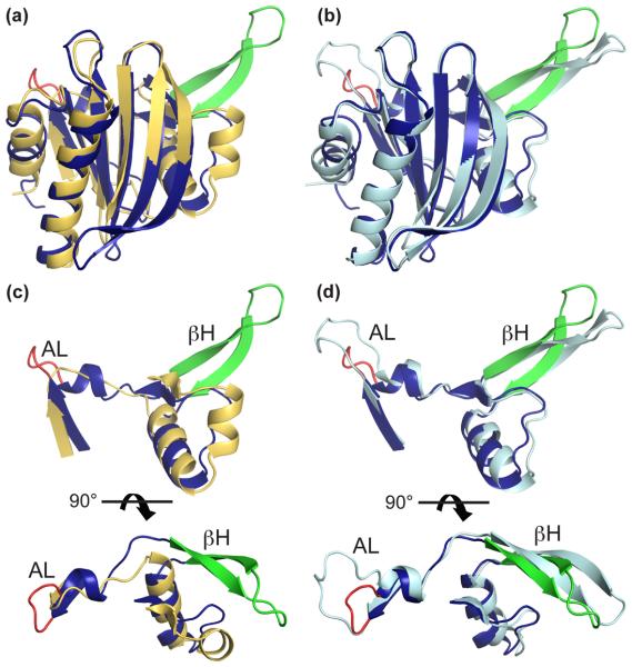
Fig. 3. Structural comparison of profilins from T. gondii, S. cerevisiae and P. falciparum.
T. gondii profilin (TgPRF) is in blue with red acidic loop and green β-hairpin. (a) Superposition of TgPRF onto S. cerevisiae profilin (PDB code 1YPR) in orange, which represents the conserved non-apicomplexan profilin structure (RMSD is 2.35 Å). (b) Superposition of TgPRF onto P. falciparum profilin (PfPRF, PDB code 2JKF) in cyan (RMSD is 1.06 Å). (c, d) The divergent features of TgPRF include a highly acidic loop (AL, TgPRF residues 37-40), and a β-hairpin (βH, residues 50-67). The latter is conserved among apicomplexans in length and overall structure, but not in sequence.

